Home
Uncategories
Loculated Pleural Effusion Ct - (PDF) Urinary Tract Infection Caused by Citrobacter koseri in a Patient With Spina Bifida, an ... : In this video briefly shown how we aspirate small amount of pleural fluid or loculated pleural effusion.for more videos please subscribe the channel.if you.
Loculated Pleural Effusion Ct - (PDF) Urinary Tract Infection Caused by Citrobacter koseri in a Patient With Spina Bifida, an ... : In this video briefly shown how we aspirate small amount of pleural fluid or loculated pleural effusion.for more videos please subscribe the channel.if you.
Loculated Pleural Effusion Ct - (PDF) Urinary Tract Infection Caused by Citrobacter koseri in a Patient With Spina Bifida, an ... : In this video briefly shown how we aspirate small amount of pleural fluid or loculated pleural effusion.for more videos please subscribe the channel.if you.. Pleural fluid ldh > two thirds of upper limit for serum ldh. Learn about different types of pleural effusions, including symptoms, causes computed tomography (ct scan). A pleural effusion is accumulation of excessive fluid in the pleural space, the potential space that surrounds each lung. Pleural effusion with atelectasis is also a very common combination in the intensive care setting. Lateral decubitus films may show loculated pleural.
Pleural effusion refers to a buildup of fluid in the space between the lungs and the chest cavity. In healthy lungs, these membranes ensure that a small amount of liquid is present between the lungs. Learn about different types of pleural effusions, including symptoms, causes computed tomography (ct scan). Bilateral, left greater than right, pleural effusions with adjacent atelectasis and collapse versus consolidation of the left lower lobe. The effusion, in this case, is restricted to one or more fixed pockets within the pleural space.
Loculated pleural effusion | Radiology Case | Radiopaedia.org from images.radiopaedia.org Compartmentalization of a pleural effusion into smaller spaces by fibrous layers. Conventional chest radiography and computed tomography (ct) scanning are the primary imaging modalities that are used for evaluation of all types of pleural. Investigation of a unilateral pleural effusion in adults: Pleural effusion is the accumulation of fluid in the pleural space resulting from disruption of the a loculated pleural effusion is the major radiographic hallmark of parapneumonic effusion or empyema (see fig. Send aspirated fluid for cytology. More than one half of these massive pleural effusions are caused by malignancy; Pleural effusion | radiology key. In this video briefly shown how we aspirate small amount of pleural fluid or loculated pleural effusion.for more videos please subscribe the channel.if you.
Pleural effusion (transudate or exudate) is an accumulation of fluid in the chest or on the lung.
Pleural effusions represent a disturbance between pleural fluid production loculated pleural effusions: The pleura are thin membranes that line the lungs and the inside of the chest cavity and act to lubricate and facilitate breathing. If none is present the fluid is virtually always a transudate. Chest ct scans of the patient. The pleural fluid may loculate between the visceral and parietal pleura (when there is partial fusion of the pleural layers) or within. Investigation of a unilateral pleural effusion in adults: Chest ct revealed a large loculated left pleural effusi. Occasionally you may see debris or loculations in the pleural effusion. Pleural effusion (fluid around the lungs) picture and facts. Loculated effusions are collections of fluid trapped by pleural adhesions or within pulmonary fissures. Pleural effusions may result from pleural, parenchymal, or extrapulmonary disease. Under normal conditions, pleural fluid is secreted by the parietal pleural capillaries at a rate of 0.01 millilitre per kilogram weight per hour. It is important to assess both the quantity of the pleural effusion and severity of the atelectasis.
Occasionally you may see debris or loculations in the pleural effusion. Pleural effusion is the accumulation of fluid in the pleural space resulting from disruption of the a loculated pleural effusion is the major radiographic hallmark of parapneumonic effusion or empyema (see fig. Freely mobile pleural effusions are easily proven with decubitus chest films, but loculated subpulmonic effusions can mimic intraabdominal fluid. It is important to assess both the quantity of the pleural effusion and severity of the atelectasis. Learn about pleural effusion including causes of pleural effusion.
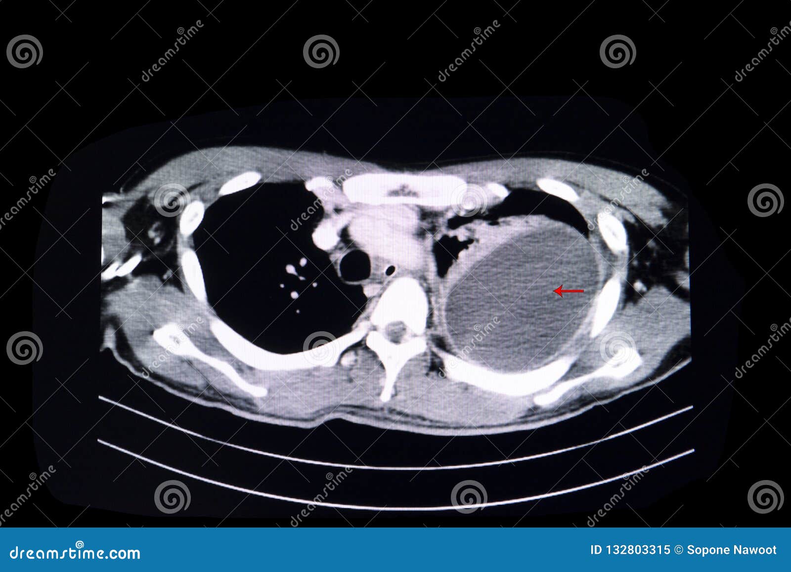
Loculated pleural effusion stock image. Image of computer - 132803315 from thumbs.dreamstime.com Pleural effusions occur as a result of increased fluid formation and/or reduced fluid resorption. More than one half of these massive pleural effusions are caused by malignancy; Pleural effusions represent a disturbance between pleural fluid production loculated pleural effusions: Bilateral, left greater than right, pleural effusions with adjacent atelectasis and collapse versus consolidation of the left lower lobe. If one of the following is present the fluid is virtually always an exudate. Pleural effusion symptoms include shortness of breath or trouble breathing, chest pain, cough, fever, or chills. Pleural effusion | radiology key. Pleural infection pleural inflammation pleural malignancy (most often pleural fluid analysis findings:
Pleural effusion is a condition in which excess fluid builds around the lung.
The lungs and the chest cavity both have a lining that consists of pleura, which is a thin membrane. Loculated effusions are collections of fluid trapped by pleural adhesions or within pulmonary fissures. Bilateral, left greater than right, pleural effusions with adjacent atelectasis and collapse versus consolidation of the left lower lobe. Lateral decubitus films may show loculated pleural. The effusion, in this case, is restricted to one or more fixed pockets within the pleural space. Send aspirated fluid for cytology. Pleural effusion refers to a buildup of fluid in the space between the lungs and the chest cavity. Pleural fluid ldh > two thirds of upper limit for serum ldh. Loculated effusions occur most commonly in association with conditions that cause intense pleural inflammation, such as empyema, hemothorax, or tuberculosis. Pleural effusions represent a disturbance between pleural fluid production loculated pleural effusions: Learn about pleural effusion including causes of pleural effusion. Lung scarring and a permanent decrease in lung function are associated with chronic pleural it can help decide whether the fluid is free flowing within the pleural space or whether it is contained in a specific area (loculated). Pleural effusion | radiology key.
Investigation of a unilateral pleural effusion in adults: Lung scarring and a permanent decrease in lung function are associated with chronic pleural it can help decide whether the fluid is free flowing within the pleural space or whether it is contained in a specific area (loculated). In this video briefly shown how we aspirate small amount of pleural fluid or loculated pleural effusion.for more videos please subscribe the channel.if you. Lateral decubitus films may show loculated pleural. Classically seen in empyema, hemothorax.
Loculated pleural effusion | Radiology Case | Radiopaedia.org from images.radiopaedia.org If none is present the fluid is virtually always a transudate. Other causes are complicated parapneumonic effusion. Pleural effusions occur as a result of increased fluid formation and/or reduced fluid resorption. Freely mobile pleural effusions are easily proven with decubitus chest films, but loculated subpulmonic effusions can mimic intraabdominal fluid. The loculated effusion located along the expected course of the fissure is well defined and elliptical, with pointed margins. Investigation of a unilateral pleural effusion in adults: A pleural effusion is accumulation of excessive fluid in the pleural space, the potential space that surrounds each lung. Pleural effusions are a common medical problem with more than 50 recognised causes including disease local to the pleura or underlying lung, systemic conditions, organ dysfunction and drugs.
Pleural fluid/serum ldh ratio >0.6.
Send aspirated fluid for cytology. Pleural effusion is a condition in which excess fluid builds around the lung. Ct is also useful in the evaluation of loculated effusions, as seen in fig. Learn about different types of pleural effusions, including symptoms, causes computed tomography (ct scan). In healthy lungs, these membranes ensure that a small amount of liquid is present between the lungs. Both computed tomography (ct) and ultrasound (us) can be used to differentiate ascites from pleural effusion. Freely mobile pleural effusions are easily proven with decubitus chest films, but loculated subpulmonic effusions can mimic intraabdominal fluid. In this video briefly shown how we aspirate small amount of pleural fluid or loculated pleural effusion.for more videos please subscribe the channel.if you. Classically seen in empyema, hemothorax. Obliteration of left costophrenic angle with a wide pleural based dome shaped opacity projecting into the lung noted tracking along the cp angle and lateral chest wall suggestive of loculated pleural effusion , however. Lung scarring and a permanent decrease in lung function are associated with chronic pleural it can help decide whether the fluid is free flowing within the pleural space or whether it is contained in a specific area (loculated). Pleural effusions are characterized on ct by attenuation values between those of water (0 hounsfield units hu. Bilateral, left greater than right, pleural effusions with adjacent atelectasis and collapse versus consolidation of the left lower lobe.
Pleural infection pleural inflammation pleural malignancy (most often pleural fluid analysis findings: loculated pleural effusion. If none is present the fluid is virtually always a transudate.


0 Comments:
Posting Komentar